Imprimir
Research
The Photobiomedical Instrumentation research group focuses on non-invasive optical techniques for monitoring vital signs and peripheral perfusion on the skin. Our goal is to extract all the potential of optical sensors and systems for non-invasive evaluations using physics tricks, aided by engineering and computational techniques. We employ traditional and neural network models to analyze the data and identify patterns. Most of our time is spent implementing and testing models and computer scripts on data and simulations, as well as thinking about physical models of light-tissue interaction and mathematical problem-solving for our applications. We occasionally collect data with volunteers.
Our physics department offers a Medical Physics bachelor’s degree, and a graduate program in Physics Applied to Medicine and Biology. Training and research includes optical methods, photobiophysics, MRI, ultrasound, radiation therapy, PET, complex systems, and computational neuroscience. Our graduate courses are taught in English, which provides an opportunity for international students to join our classroom. In addition, we have exchange programs with leading American, European, and Asian universities, which further enhances the collaborative nature of our research.
We are based at the University of São Paulo (USP) in Ribeirão Preto, which is renowned for its research in the biological and medical fields. Ribeirão Preto is also a national hub for the medical-pharmaco-dental industry. Our affiliation with USP’s government-sponsored synergistic startup incubator provides opportunities for synergetic collaborations with this industry.
Full fellowships are available for all qualified Master’s and Ph.D. candidates, regardless of their nationality. Entrance exams can be arranged to be taken abroad at a local university and, upon explicit request, can be taken in English.
Current subprojects with potential for Masters and PhD projects:
- Quantitative measurements using optical and video imaging, such as determining the degree of radiodermatitis through polarized light images.
- Optical spectroscopy for developing spectral separation methods to identify compounds (e.g., drugs) or patterns characterizing states such as cancer, inflammation, and normal tissue.
- Determining vital signs and physiological parameters through optical instrumentation, including the Magic Mirror for qualitative visualization of blood perfusion and techniques for measuring blood flow velocity using video.
- Developing image processing systems using cameras and polarized light for augmented visualization in medical and dental clinical applications.
- Scaling of epidemic thresholds and individual risk with population size for discrete agents
- Non-invasive or remote camera-based measurements of blood pressure.
- Early detection of retinopathies, which can potentially diagnose diabetes, Alzheimer’s, cognitive impairment, glaucoma, and others, is achieved using image oximetry. This technique uses a video camera and spectral image processing. (In collaboration with the Ophthalmology Department, USP-FMRP)
Mathematical tools used in the projects include:
- Machine Learning (Neural Networks, Old-style Regression with Physical Models)
- Descriptive Statistics
- Linear Algebra (Matrix Algebra, ICA, PCA, etc.)
Physical Tools and Systems, include:
- Spectroscopy (Absorption/Transmission, Visible and Near-Visible)
- Colorimetry (CIELAB) and Color Calibration
- Cameras (RGB Sensors), photodiodes and other sensors and systems.
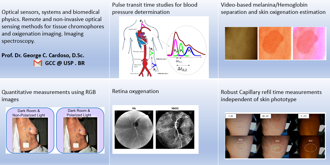
Our research applications include analyzing blood perfusion, pulse transit time, determining non-occlusive blood pressure, measuring capillary filling time, assessing tissue oxygenation, and quantifying and mapping melanin in the skin. Additionally, we collaborate with the school of medicine to explore Optical Coherence Tomography and retinography images, which utilize the physics of light-tissue interactions and have the potential to predict ocular and brain diseases at an early stage.
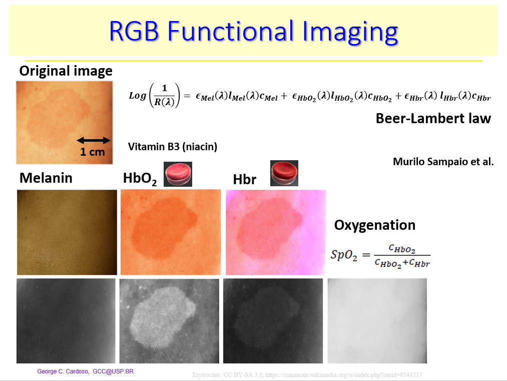
Showcase:
The images below illustrate some of our studies that utilize multispectral spectroscopy in conjunction with RGB camera channels to gather spatial, spectral, and temporal information about both biological and non-biological systems. By combining optical techniques with image processing, we are able to produce images in which different skin chromophores, such as melanin and hemoglobin, have been separated.