
Tissue Characterization
Elastography
Elastography is a non-invasive and painless medical imaging technique that quantitatively explores the elastic properties and stiffness of tissues. Its objective is to provide diagnostic information regarding the presence or status of diseases by assessing tissue stiffness. For example, cancerous tumors typically exhibit greater stiffness compared to the surrounding healthy tissues. By evaluating tissue elasticity, elastography facilitates the diagnosis of conditions affecting various organs such as the liver, breast, thyroid, kidneys, among others. Additionally, it furnishes anatomical images for comparison and can be integrated with other techniques to enhance clinical treatments.
The main steps in elastography encompass the following:
1 – Application of either static or dynamic force in the region of interest. This introduces a disturbance, typically a mechanical wave, into the tissue under analysis. Various methods can be employed for this purpose, such as ultrasonic radiation force, magnetic force, or laser-based ultrasound, among others.
2 – Observation of the tissue’s response to the applied disturbance. This involves the use of tools like an ultrasonic transducer to detect variations in the amplitude and phase of the traveling wave.
3 – Determination of mechanical properties (i.e., stiffness) based on the observed response. For example, stiffer tissues tend to propagate waves faster and, hence, the higher the elastic modulus of the medium. Therefore, characterizing tissue involves analyzing factors like time delay, energy content of waves, and dispersion curves, among others.
4 – Reporting of results, which entails displaying the formed image along with corresponding deformation, velocity or stiffness maps.
Acoustography
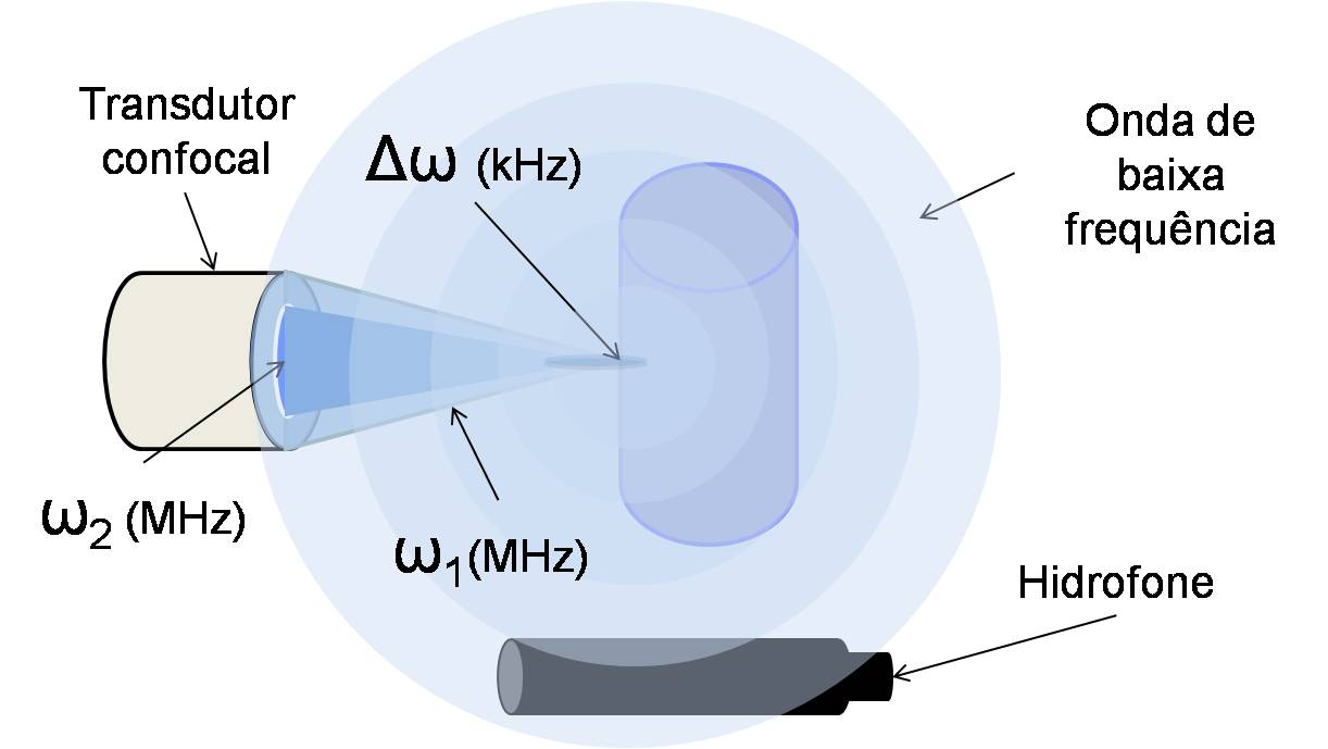
Acoustography is a non-invasive quantitative ultrasonic technique that generates speckle-free images by leveraging the medium’s response to harmonic radiation force induced by ultrasound. Acoustography doesn’t rely on ionizing radiation. Instead, it uses ultrasound energy to produce near real-time images. Moreover, this technique can visualize features within materials that may be opaque or exhibit high absorption of electromagnetic waves. Acoustography capitalizes on the non-linear characteristics of acoustic propagation, where energy converts from high frequencies (typically MHz) to low frequencies (typically kHz). For example, for tissue characterization, ultrasound radiation force is used to excite tissues at a low frequency, and the corresponding acoustic response is then used to generate images, providing insight into the mechanical properties and potential abnormalities within the tissue. Biomedical applications of acoustography encompass the assessment of various organs such as the breast, prostate, arteries, liver, and thyroid, among others.
The main steps in acoustography consist of:
1 – Ultrasound energy conversion (non-linear behavior), involving the use of high-frequency ultrasound energy to elicit a low-frequency response in the tissue. Typically, this is achieved by directing two high-frequency ultrasound beams with slightly different frequencies toward the tissue.
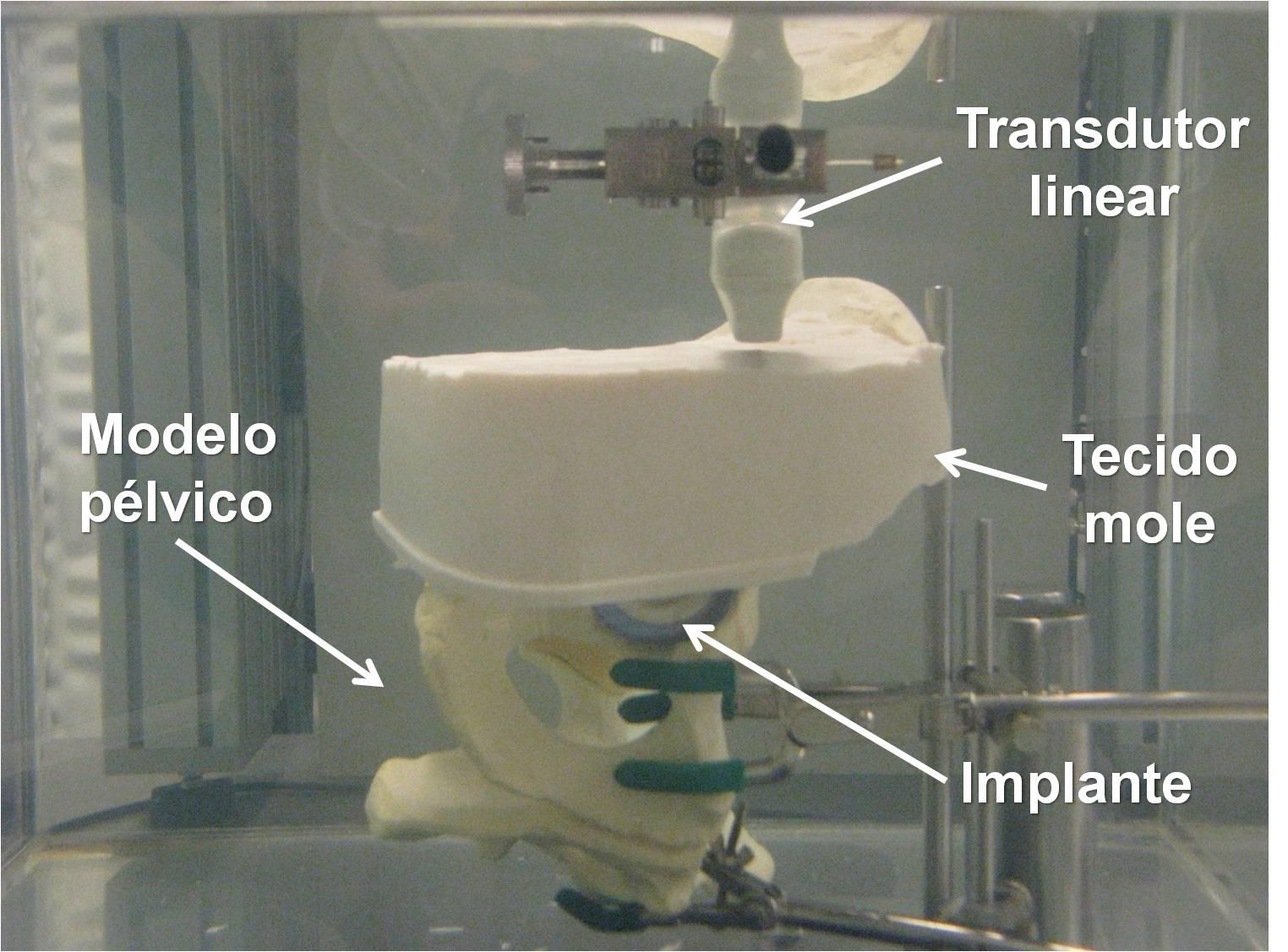
2 – Detection, which entails receiving the low-frequency energy using either a single or multi-element transducer.
3 – Image formation, where the detected sound waves are used to create an image highly sensitive to the stiffness of the medium. This sensitivity makes acoustography valuable for detecting abnormalities such as fibrosis and tumors. Furthermore, the absence of speckle noise in the obtained images enhances clarity and visualization.

David Alejandro
Postdoctoral Fellow